 RadWorld
Classification
Improve chest abnormality classification with synthetic training scans.
Pretrained on 232,781 curated scans from 152 datasets across 23 countries, RadWorld learns a shared generative prior across anatomical regions and imaging modalities.
CT and MRI tasks differ in the information available and the examination required. RadWorld brings scan generation, imaging translation and treatment simulation into a common formulation: generate a target 3D volume from the supplied conditions.
Generation
Generate CT and MRI volumes, and guide CT synthesis with radiology reports or segmentation masks.
Translation
Condition on an available scan to generate a target modality, MRI sequence or contrast phase.
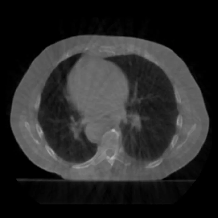
Cone-beam CT
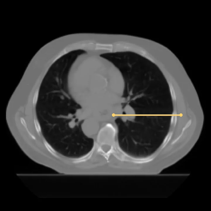
CT
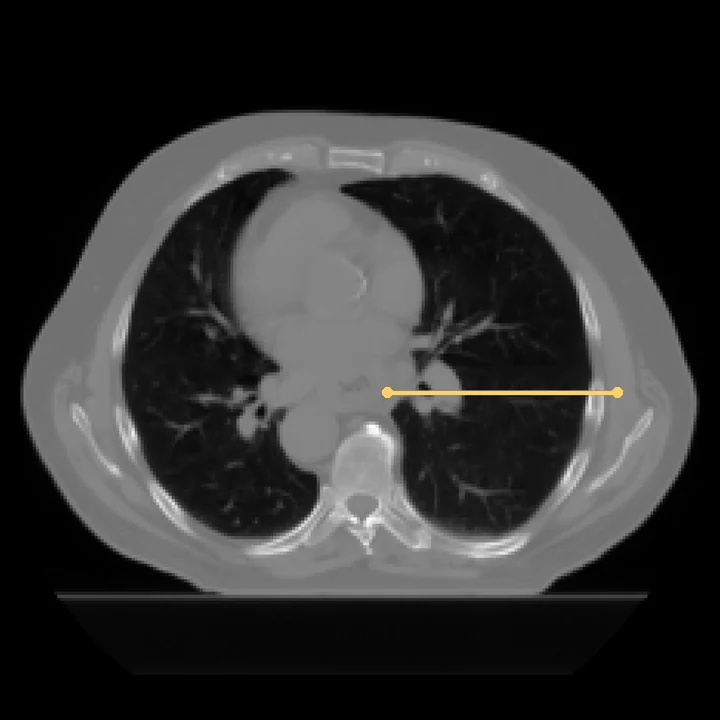
CT
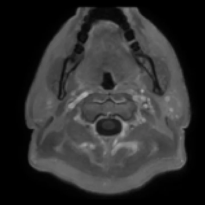
MRI
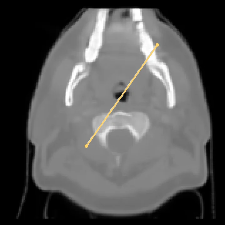
CT
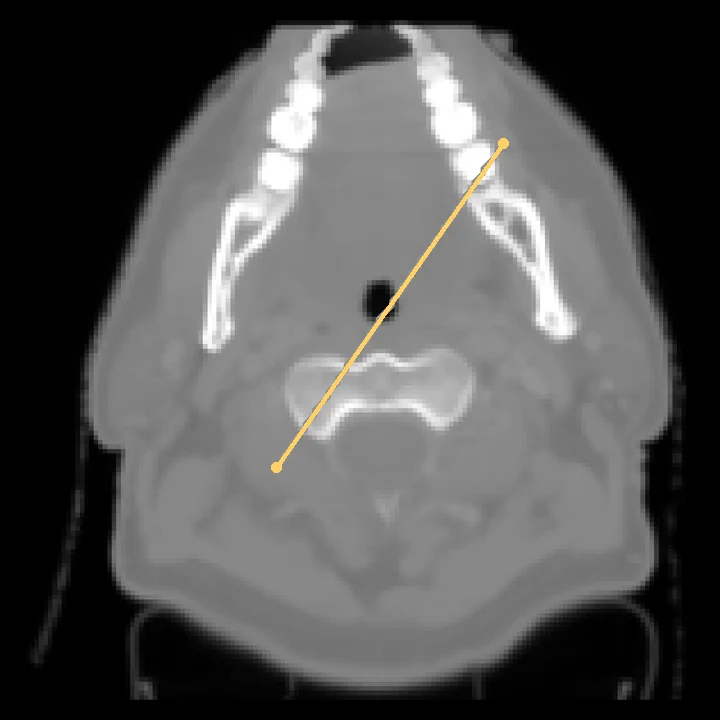
CT
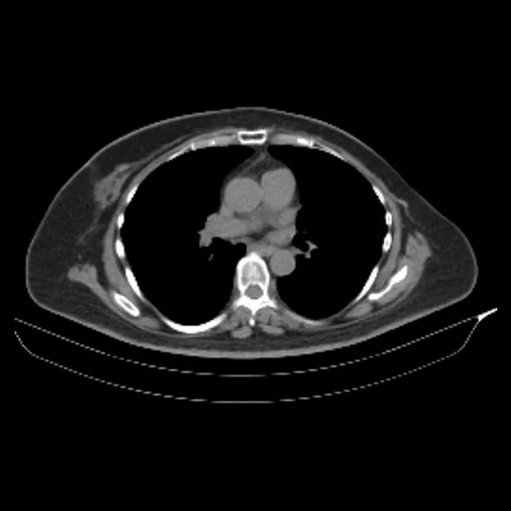
Non-contrast CT
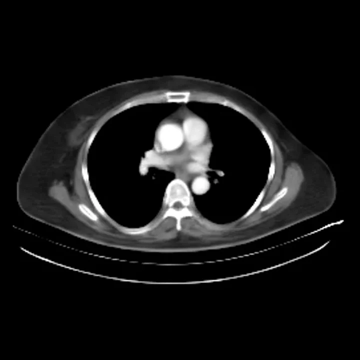
Arterial-phase CT
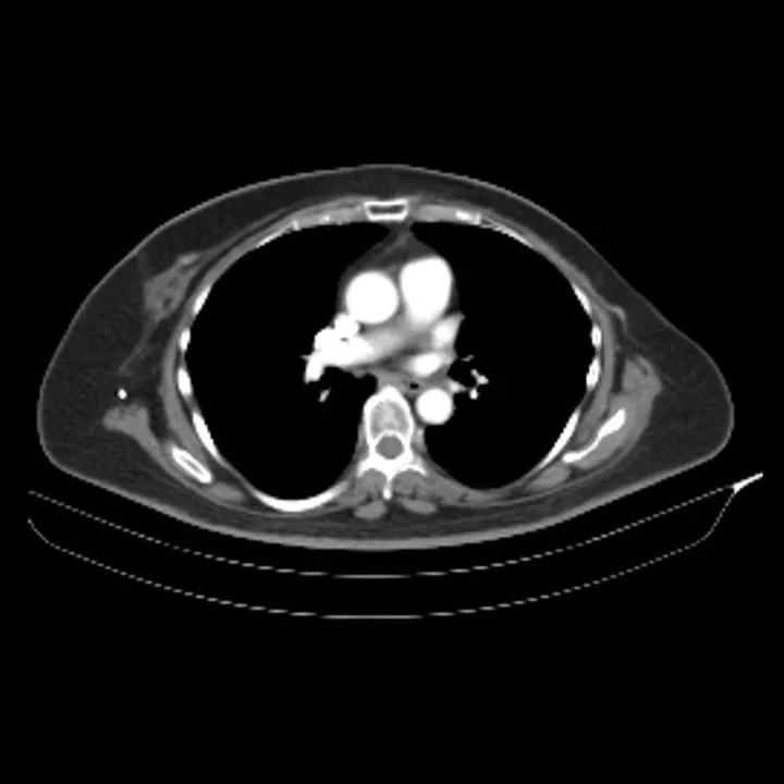
Arterial-phase CT
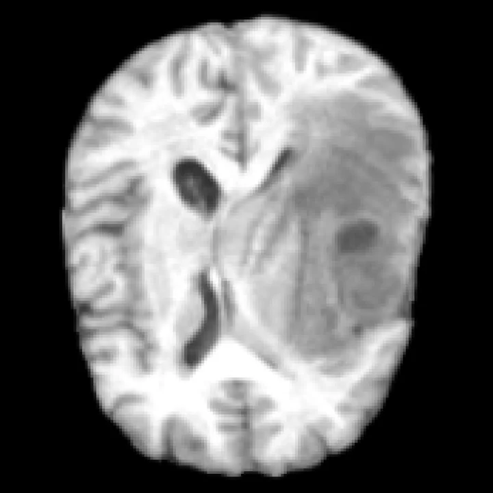
T1 + T2 + FLAIR
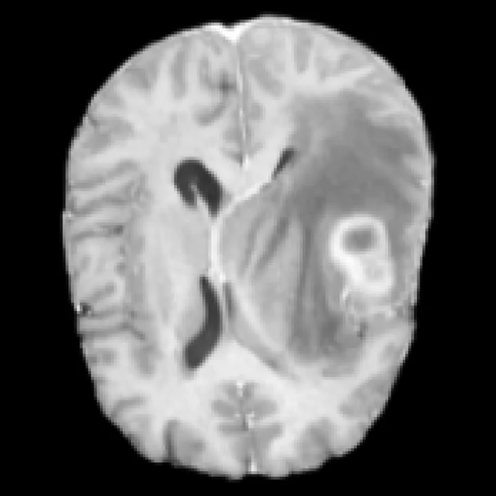
T1CE MRI
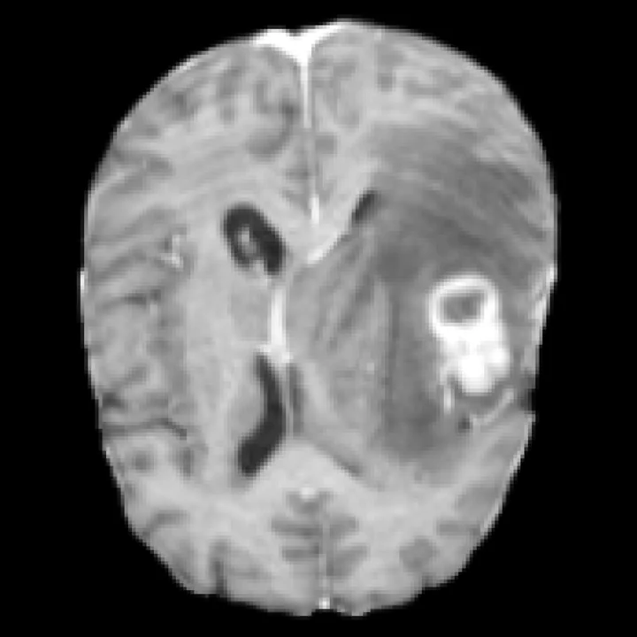
T1CE MRI
Simulation
Condition on pre-treatment CT and TACE details to generate virtual post-treatment scans for hepatocellular carcinoma.
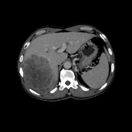
CT before TACE
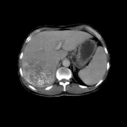
Generated by RadWorld
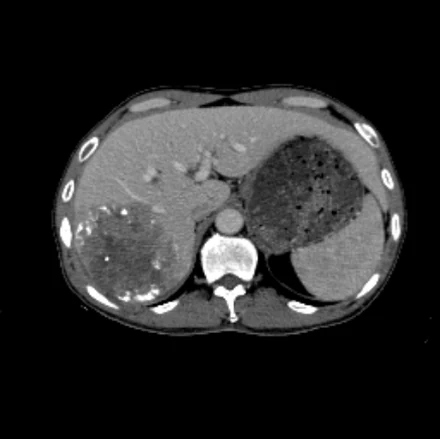
Real CT after TACE
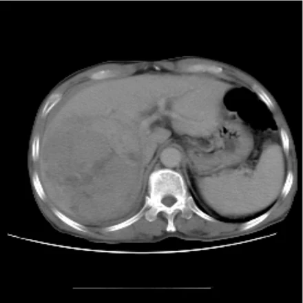
CT before TACE
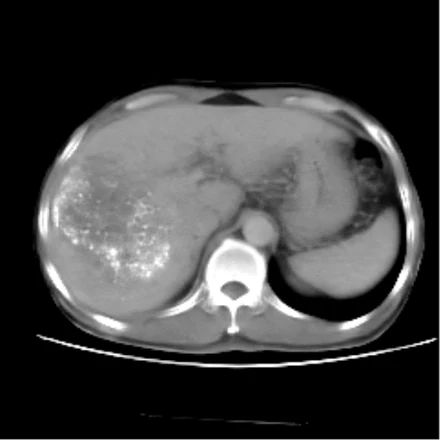
Generated by RadWorld
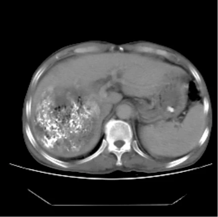
Real CT after TACE
Visual Turing test
Explore ten tumor scans and decide which are real and which are generated. View the radiologist reader study to see how experts performed.
Your answers stay in this browser.
Downstream applications
Use RadWorld-generated scans to augment training data for classification, segmentation and report generation, or as inputs for survival prediction.
Improve chest abnormality classification with synthetic training scans.
Improve segmentation across eight tumor types with synthetic training examples.
Train report generation models with paired synthetic scans and reports.
Use virtual post-TACE CT to support progression-free survival prediction.
Synthetic-to-real training ratio: 2:1.
AUC for chest CT abnormalities, higher is better
| Abnormality | Real data only | + GenerateCT | + MedSyn | + RadWorld |
|---|---|---|---|---|
| Hiatal hernia | 0.477 | 0.483 | 0.489 | 0.609 |
| Lung opacity | 0.546 | 0.559 | 0.613 | 0.632 |
| Atelectasis | 0.566 | 0.582 | 0.608 | 0.690 |
| Adenopathy | 0.623 | 0.612 | 0.623 | 0.650 |
| Pericardial effusion | 0.633 | 0.641 | 0.635 | 0.672 |
| Calcification | 0.579 | 0.614 | 0.680 | 0.688 |
| Lung nodule | 0.609 | 0.647 | 0.673 | 0.673 |
| Emphysema | 0.535 | 0.586 | 0.676 | 0.772 |
| Bronchiectasis | 0.610 | 0.679 | 0.615 | 0.784 |
| Consolidation | 0.644 | 0.657 | 0.724 | 0.723 |
| Fibrotic sequela | 0.645 | 0.756 | 0.737 | 0.826 |
| Pleural effusion | 0.790 | 0.813 | 0.852 | 0.902 |
| Overall | 0.613 | 0.650 | 0.665 | 0.723 |
Synthetic-to-real training ratio: 2:1.
Dice for eight cancers, higher is better
| Cancer | Real data only | + DiffTumor | + FreeTumor | + RadWorld |
|---|---|---|---|---|
| NPC | 0.576 | 0.590 | 0.598 | 0.629 |
| HNSCC | 0.632 | 0.653 | 0.654 | 0.675 |
| NSCLC | 0.625 | 0.651 | 0.691 | 0.723 |
| HCC | 0.599 | 0.700 | 0.703 | 0.730 |
| PDAC | 0.487 | 0.541 | 0.535 | 0.588 |
| CRC | 0.438 | 0.476 | 0.476 | 0.531 |
| RCC | 0.729 | 0.762 | 0.764 | 0.783 |
| BLCA | 0.673 | 0.726 | 0.724 | 0.764 |
Synthetic-to-real training ratio: 2:1.
Text similarity and clinical accuracy, higher is better
| Region | Real data only | + GenerateCT | + MedSyn | + RadWorld |
|---|---|---|---|---|
| Chest | 0.215 | 0.263 | 0.264 | 0.270 |
| Abdomen | 0.320 | 0.340 | 0.342 | 0.358 |
| Pelvis | 0.193 | 0.252 | 0.250 | 0.258 |
| Chest | 0.311 | 0.320 | 0.316 | 0.324 |
| Abdomen | 0.413 | 0.426 | 0.429 | 0.442 |
| Pelvis | 0.373 | 0.377 | 0.372 | 0.388 |
| Chest | 0.322 | 0.336 | 0.339 | 0.352 |
| Abdomen | 0.373 | 0.399 | 0.395 | 0.410 |
| Pelvis | 0.424 | 0.428 | 0.429 | 0.435 |
| Chest | 0.266 | 0.274 | 0.288 | 0.314 |
| Abdomen | 0.421 | 0.442 | 0.446 | 0.501 |
| Pelvis | 0.665 | 0.686 | 0.690 | 0.741 |
Progression-free survival after TACE, evaluated in two cohorts.
C-index for progression-free survival, higher is better
| Cohort | Pre-TACE CT only | + patient and procedural variables | + real post-TACE CT | + virtual post-TACE CT |
|---|---|---|---|---|
| Cohort A | 0.577 | 0.595 | 0.732 | 0.727 |
| Cohort B | 0.571 | 0.595 | 0.726 | 0.721 |
Progression-free survival of risk groups predicted from virtual post-TACE CT
Hazard ratio 2.73, P < 0.0001
Progression-free survival of risk groups predicted from virtual post-TACE CT
Hazard ratio 2.45, P < 0.0001
Research team
‡ Equal contribution · * Corresponding authors
1Monash University 2Ant Healthcare 3Shanghai Artificial Intelligence Laboratory 4Stanford University School of Medicine 5Shanghai Innovation Institute 6The First Affiliated Hospital of Jinan University 7Fudan University 8Fuzhou University
Resources
@article{ye2026radworld,
  title   = {A World Foundation Model for 3D Tomographic Medical Imaging},
  author  = {Ye, Jin and Ma, Chenglong and Zhang, Lu and others},
  journal = {arXiv preprint},
  year    = {2026}
}